Mechanism of Action and Principal Research Findings
An ordered reading of the BPC-157 literature from molecular pathway to multi-system preclinical evidence.
- [01] Understand the primary molecular pathways through which BPC-157 exerts its effects in animal models.
- [02] Identify the tissue systems where preclinical evidence is strongest and where it is more limited.
- [03] Distinguish between in vitro findings, rodent in vivo findings, and the limited human pilot data.
- [04] Understand why BPC-157's angiogenic effect is described as tissue-context-dependent rather than uniformly pro-angiogenic.
Mechanism of Action
The primary signaling nodes through which BPC-157 exerts its tissue-repair effects across dozens of rodent studies.
 F1
F1
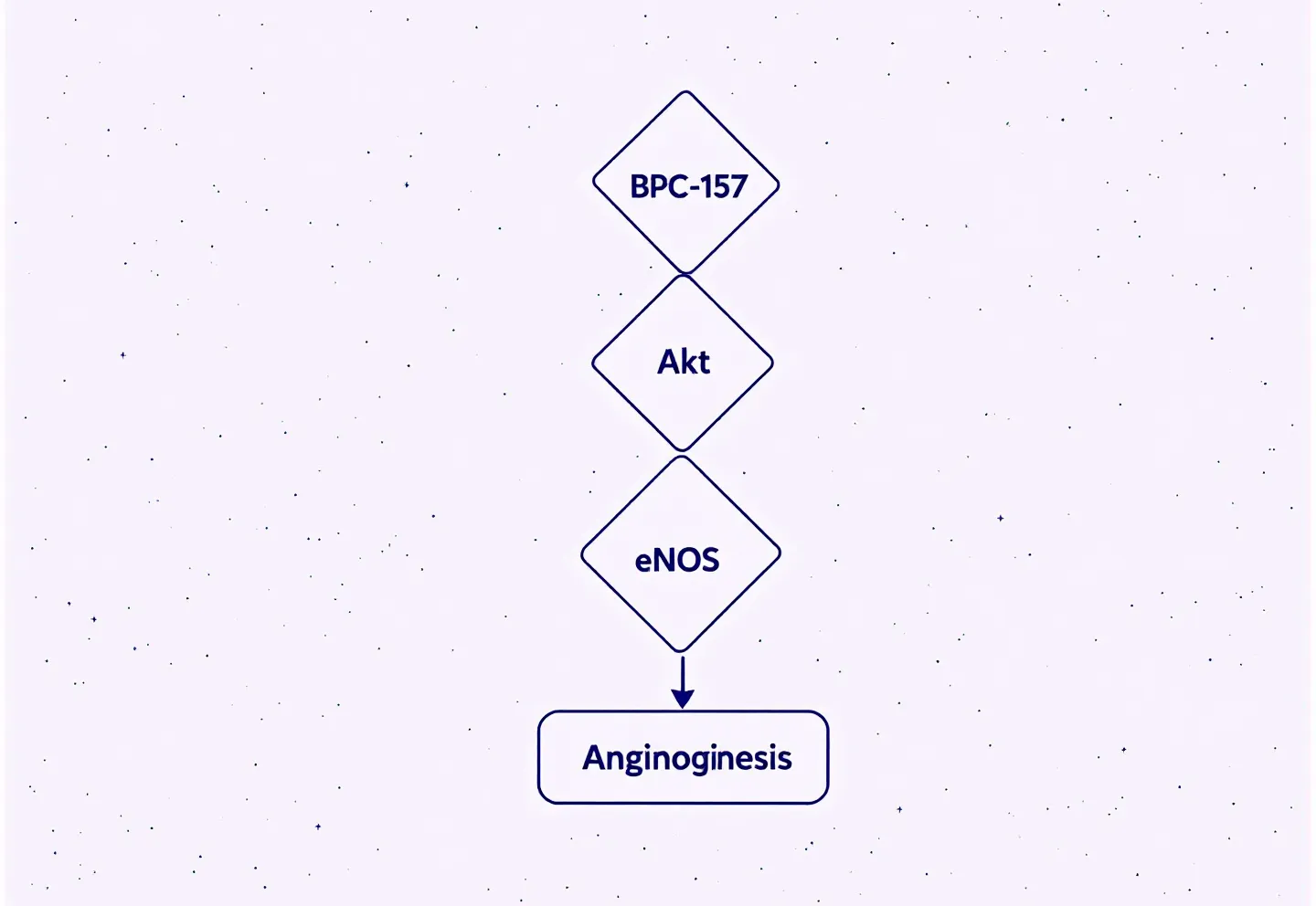
FIG. 01 — BPC-157 mechanistic cascade: VEGFR2 upregulation driving the Akt-eNOS axis to tissue-adaptive angiogenesis.
BPC-157 is a 15-amino-acid peptide (GEPPPGKPADDAGLV, MW 1419.5 Da) derived from a protein fraction in human gastric juice. Its mechanistic signature across dozens of rodent studies can be organized around four primary signaling nodes.
VEGFR2 upregulation. Vascular endothelial growth factor receptor 2 is a cell-surface kinase that, when activated, drives endothelial cell proliferation and migration — the fundamental cellular machinery of angiogenesis. BPC-157 consistently upregulates VEGFR2 expression in healing tissue, and this appears to be the primary upstream event governing its pro-angiogenic activity in tendon and muscle repair models.[1][4]
Akt-eNOS axis activation. Downstream of VEGFR2, Akt phosphorylates endothelial nitric oxide synthase (eNOS), stimulating nitric oxide production. Nitric oxide drives vasodilation, promotes endothelial survival, and contributes to local angiogenesis. Elevated Akt1 and Nos3 mRNA are among the genes most consistently upregulated in BPC-157 studies — Seiwerth et al. (2021) documented upregulation of both within 10 minutes of BPC-157 application to wound tissue in rats.[4]
ERK1/2 and FAK-paxillin pathway. Focal Adhesion Kinase (FAK) and its adaptor protein paxillin form a signaling complex that mediates fibroblast adhesion, migration, and proliferation. ERK1/2 is a downstream kinase that regulates collagen synthesis. BPC-157 activates both pathways, which accounts for its effects on fibroblast activity in tendon cultures. Chang et al. (2014) demonstrated that at concentrations of 0.1–0.5 μg/mL in isolated rat Achilles tendon fibroblasts, BPC-157 produced up to sevenfold increases in growth hormone receptor mRNA and protein by day three of culture — and enhanced JAK2 phosphorylation (a downstream GH signaling event) when GH was subsequently introduced.[3]
NF-kB downregulation and nitric oxide modulation. BPC-157 reduces NF-kB activity, a master regulator of inflammatory cytokine production. Concurrently, it modulates the full nitric oxide system — increasing eNOS-derived NO (vasodilatory, pro-healing) while decreasing iNOS-derived NO (inflammatory, tissue-damaging). In the hippocampal ischemia model, Vukojevic et al. (2020) observed elevated Egr1, Akt1, Vegfr2, and Nos3 expression with simultaneous decreases in Nos2 (iNOS) and NF-kB.[5]
BPC-157's angiogenic behavior is not uniformly pro-angiogenic. In tendon and muscle healing models it promotes neovascularization correlated with improved repair. In corneal neovascularization models and liver cirrhosis models, BPC-157 opposes pathologic angiogenesis.[11] The 2025 Sikiric et al. review describes this as a "beneficial pleiotropic effect" — context-adaptive rather than constitutively stimulatory.
Musculoskeletal Findings
The most extensively documented area of BPC-157 preclinical research, spanning tendon-to-bone healing, muscle-to-bone reattachment, and in vitro fibroblast activity.
 F2
F2
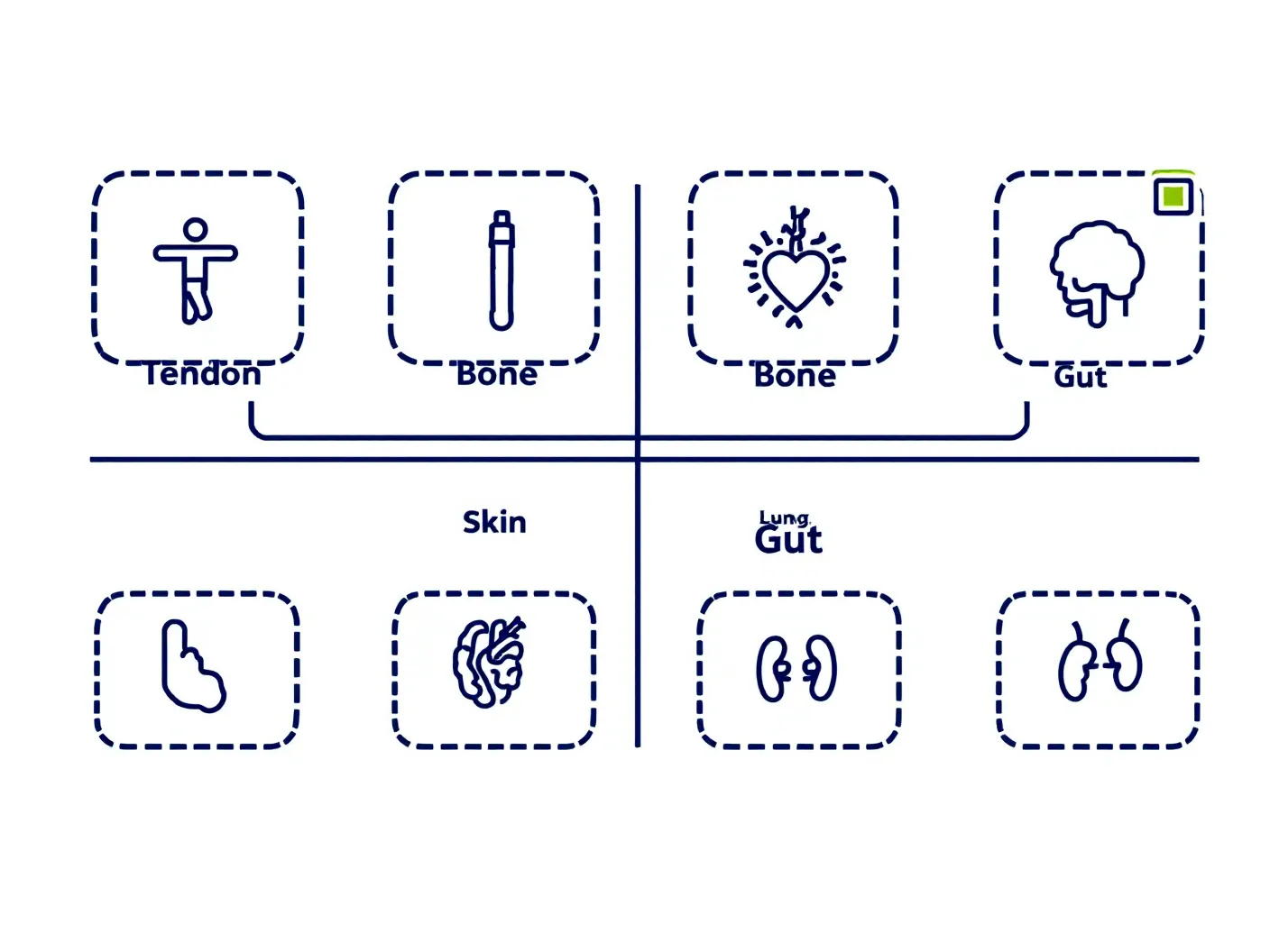
FIG. 02 — Tissue systems studied in BPC-157 preclinical literature; green markers indicate the three with the strongest multi-study evidence base (tendon, bone, gut).
Achilles tendon-to-bone healing. Krivic et al. (2006) studied BPC-157 in rats following surgical detachment of the Achilles tendon. Intraperitoneal administration at doses spanning three orders of magnitude — 10 μg/kg, 10 ng/kg, and 10 pg/kg — consistently produced superior load-to-failure values, stiffness, and Young's modulus compared to controls. Importantly, BPC-157 also counteracted the healing impairment induced by methylprednisolone (a corticosteroid known to degrade tendon quality), restoring Achilles functional index scores and collagen fiber organization when administered alongside or after the corticosteroid.[2] PRECLINICAL
Angiogenesis in crushed and transected tissue. Brcic et al. (2009) examined BPC-157 in rat models of both crushed muscle and transected muscle/tendon. Immunohistochemical analysis showed upregulated VEGF expression and more organized angiogenesis at the repair site compared to controls. Notably, BPC-157 showed no direct angiogenic effect in cell culture in the same study — its activity required an in vivo healing environment, suggesting the mechanism is not simple VEGF induction but is dependent on the tissue signaling context.[1] PRECLINICAL
Growth hormone receptor in tendon fibroblasts. Chang et al. (2014) isolated Achilles tendon fibroblasts from rats and exposed them to BPC-157 at 0.1–0.5 μg/mL. Growth hormone receptor expression increased dose-dependently and time-dependently, reaching up to sevenfold increases in mRNA and protein by day three. Adding GH to BPC-157-treated cells produced significantly greater JAK2 phosphorylation than GH alone — a finding the authors interpret as receptor sensitization.[3] IN VITRO
Quadriceps muscle-to-bone reattachment. Matek et al. (2025) studied oral BPC-157 at 10 μg/kg or 10 ng/kg following surgical detachment of the quadriceps muscle from its attachments in rats. Treatment was given by oral gavage five minutes post-surgery and then in drinking water. At 90 days, treated animals showed consistent muscle-to-bone reattachment with mature, parallel-oriented collagen fibers and organized bone at the interface. Control animals showed permanent knee flexure contracture and inability to extend the limb.[12] PRECLINICAL
Gastrointestinal Findings
BPC-157's origins in gastric cytoprotection research, and the extension of gastrointestinal findings to wound healing, fistula closure, and vascular occlusion.
Wound healing across skin and gut. Seiwerth et al. (2021) published a comprehensive wound-healing study covering skin incisions, burns, diabetic ulcers, and fistulas in rats. BPC-157 was administered via three distinct routes — topical (1 μg/g cream), intraperitoneal (10 μg/kg down to 10 pg/kg), and oral gavage. Across all models and all routes, BPC-157 accelerated re-epithelialization, enhanced angiogenesis, increased tensile strength of healed tissue, and outperformed silver sulfadiazine in the burn model. Gene expression analysis documented upregulation of 19 genes — including Akt1, VEGFA, Egr1, and Nos3 — within 10 minutes of application.[4] PRECLINICAL
Fistula closure. Vukusic et al. (2024) created experimentally induced duodenocolic fistulas in Wistar rats and treated them with BPC-157 at 10 μg/kg and 10 ng/kg via local application, intragastric gavage, oral administration, and intraperitoneal injection. All routes produced complete closure of both the duodenal and colonic defects, elimination of fistula leakage, prevention of weight loss, and significantly reduced adhesion formation compared to untreated controls.[15] PRECLINICAL
Vascular occlusion and organ protection. Sikiric et al. (2022) examined BPC-157 across four models of major vessel occlusion: inferior caval vein occlusion, portal vein occlusion, Pringle maneuver-induced ischemia-reperfusion, and Budd-Chiari syndrome. In each model, BPC-157 rapidly recruited collateral vessels, reversed portal and caval hypertension, prevented thrombosis, and attenuated lesions across the liver, kidney, gastrointestinal tract, and heart.[9] PRECLINICAL
Neurological and Cardiovascular Findings
BPC-157 in ischemia-reperfusion, spinal cord compression, dopaminergic modulation, pulmonary hypertension, and remote organ protection models.
Hippocampal ischemia-reperfusion. Vukojevic et al. (2020) clamped bilateral carotid arteries in rats, producing hippocampal ischemia. Local BPC-157 application at 10 μg/kg — applied as a bath to the carotid triangle — produced complete functional recovery at 24 and 72 hours on Morris Water Maze, beam-walking, and lateral push tests. Histopathology showed far fewer ischemic neurons in treated animals. Gene expression analysis confirmed elevated Egr1, Akt1, Vegfr2, and Nos3 with decreased Nos2 and NF-kB.[5] PRECLINICAL
Spinal cord compression. Perovic et al. (2019) administered a single intraperitoneal injection of BPC-157 at either 200 μg/kg or 2 μg/kg within 10 minutes of spinal cord compression in rats. Both dose levels produced progressive motor recovery, resolution of spasticity by day 15, and prevention of autotomy through 360 days of observation. Controls showed persistent debilitation. Histopathology confirmed reduced vacuole formation and motoneuron degeneration in treated animals.[6] PRECLINICAL
Dopaminergic system. Vukojevic et al. (2022) reviewed BPC-157's effects across multiple rat models of dopamine system disturbance — receptor blockade, receptor supersensitivity, nigrostriatal damage, and vesicle depletion. BPC-157 normalized dopaminergic signaling and reversed haloperidol-induced catalepsy. The review proposes that peripheral administration achieves central nervous system effects via the nitric oxide system.[7] REVIEW
Pulmonary arterial hypertension. Udovicic et al. (2021) used monocrotaline-induced pulmonary arterial hypertension in rats. BPC-157 at 10 μg/kg or 10 ng/kg, given intraperitoneally or in drinking water, both prevented and reversed pulmonary arterial wall thickening, right ventricular hypertrophy, and abnormal QT intervals. Untreated controls showed 50% mortality; treated groups showed 0% mortality.[8] PRECLINICAL
Remote organ protection. Demirtas et al. (2025) examined organ damage distant from the injury site after 45-minute lower-extremity ischemia in rats followed by 2-hour reperfusion. A single 20 μg/kg intraperitoneal dose significantly attenuated glomerular vacuolization, tubular dilation, alveolar congestion, and hepatocyte degeneration in kidney, lung, and liver respectively. Total antioxidant status improved and oxidative stress index decreased in treated animals.[14] PRECLINICAL
Human pilot data. The 2025 narrative review by McGuire et al. identified three published human studies.[13] First: intra-articular knee injection in 14 patients, with 87.5% reporting significant pain relief. Second: intravesical injection for interstitial cystitis in 12 patients, with 80–100% symptom resolution. Third: an IV safety study in 2 healthy volunteers at doses up to 20 mg, with no adverse events and no clinically meaningful laboratory changes. The authors explicitly characterize these as preliminary pilot data and note that no Phase 2 or Phase 3 randomized controlled trials have been completed. HUMAN PILOT